Picture Of Forearm Muscles And Tendons - Body Anatomy: Upper Extremity Tendons | The Hand Society - Cross sectional anatomy of the upper limb :
bymamazapatero-
0
Picture Of Forearm Muscles And Tendons - Body Anatomy: Upper Extremity Tendons | The Hand Society - Cross sectional anatomy of the upper limb :. If you keep your hand flat on a table and. Also, pollicis means thumb in latin. There are many muscles in the forearm. The muscles of the upper arm are responsible for the flexion and extension of the forearm at the elbow joint. The muscles of the forearm are predominantly slow twitch. slow twitch muscles are very resistant alternate days so that the muscles and tendons have time to recover from the previous workout.
The muscle fibers then descend towards the wrist area where they converge onto a narrow tendon. Tendons are attached to muscles and to bone. Grade i strain of forearm muscle: The thorough and detailed descriptions helped, and definitely the pictures. Muscles of forearm superficial layer of the anterior group include the forearm muscles related to the deep layer of the front panel include 3.
Medical anatomy, Hand therapy, Muscle anatomy from i.pinimg.com The extensor digitorum is a muscle belly, passing first into four tendons, which in turn transformirovalsya in stretching the tendon fixed to the base of the. The muscle fibers then descend towards the wrist area where they converge onto a narrow tendon. If you keep your hand flat on a table and. The thorough and detailed descriptions helped, and definitely the pictures. Cross sectional anatomy of the upper limb : Posterior distal shaft of ulna and iterosseous membrane i: Most of these originate from the lateral epicondyle. Do it yourself as shown in the picture!
The muscle fibers then descend towards the wrist area where they converge onto a narrow tendon.
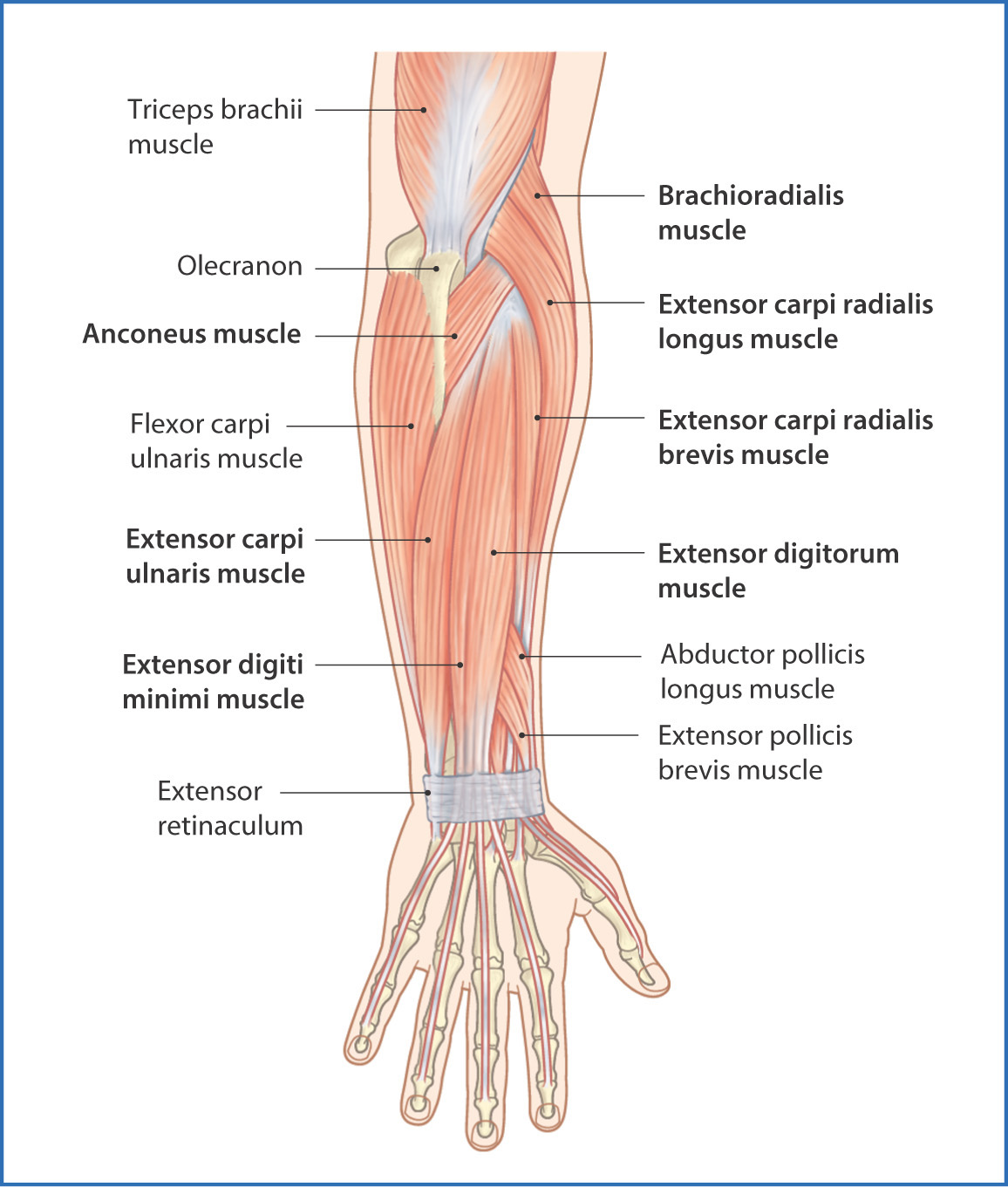
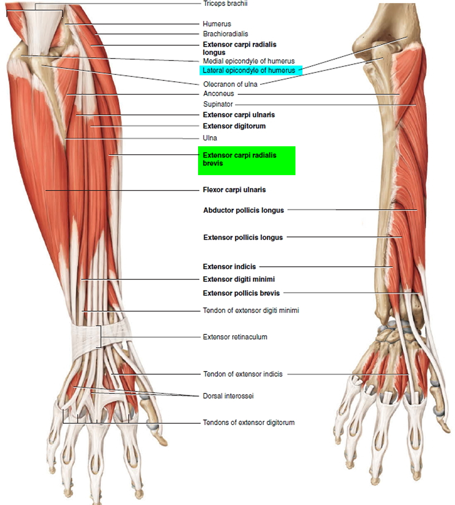
From superior to inferior, origin. See anatomy pictures of the 27 bones in the hand and wrist, how they are connected with tendons and muscles and the nerves that run through the skeletal structure. A few remaining muscles for our skeletons. Most of these originate from the lateral epicondyle. Tendons are attached to muscles and to bone. The tendons travel down the forearm through a tough band of tissue on top of the wrist. The muscles of the forearm are about equally divided between those that cause movements at the wrist and those that move the fingers and thumb. Slip to the edc tendon is responsible for flexing the mcp joint and simultaneously. Learn vocabulary, terms and more with flashcards, games and other study tools. The muscle fibers then descend towards the wrist area where they converge onto a narrow tendon. The muscles of this group take origin from the medial epicondyle of the humerus by a common tendon; In the anterior compartment, they are split into three categories: They receive additional fibers from the deep fascia of the forearm near the elbow, and from the septa which pass from this fascia between the individual muscles.
Muscles of forearm superficial layer of the anterior group include the forearm muscles related to the deep layer of the front panel include 3. Grade i strain of forearm muscle: The extensor carpi ulnaris muscle is the most medial muscle in the superficial posterior compartment of the forearm. The term forearm is used in anatomy to distinguish it from the arm. Originates from the anterior surface of the ulna and attaches to the.
posterior forearm muscles from basicmedicalkey.com Slip to the edc tendon is responsible for flexing the mcp joint and simultaneously. The longer the muscles in the forearm are (and therefore the shorter their tendons are), the easier it will be to develop them. Forearm muscle anatomy forearm muscles hand bone anatomy images hypermobility anatomy for artists chinese medicine human anatomy physical foot anatomy human anatomy forearm muscle anatomy peroneus longus human muscular system ligaments and tendons human leg human. By moving the mouse cursor over a particular area of the arm or forearm, this area is highlighted and the labels are displayed: The tendons of these muscles pass through a small corridor in the wrist known as the carpal tunnel. The tendons travel down the forearm through a tough band of tissue on top of the wrist. The term forearm is used in anatomy to distinguish it from the arm. Posterior distal shaft of ulna and iterosseous membrane i:
Do it yourself as shown in the picture!
Anterior, lateral or posterior compartment. The muscle fibers then descend towards the wrist area where they converge onto a narrow tendon. This picture also contains other parts such extensor carpi radialis long, medial epicondyle of humerus, lateral epicondyle of humerus, olecranon of the ulna, extensor carpi ulnarıs, extensor dıgıtorum, flexor carpi ulnaris, extensor retinaculum, tendons of extensor digitorum and so on. A deep layer, intermediate layer and superficial layer. Tusindvis af nye billeder af høj kvalitet tilføjes hver dag. There are many muscles in the forearm. The anterior forearm muscles are divided into 3 muscular layers; Tendon of extensor digitorum at 2nd metacarpal. In the anterior compartment, they are split into three categories: Tendons are attached to muscles and to bone. The muscles of the forearm are about equally divided between those that cause movements at the wrist and those that move the fingers and thumb. The muscles of this group take origin from the medial epicondyle of the humerus by a common tendon; Forearm muscles in the anterior compartment are arranged in superficial, intermediate and deep categories.
The muscles of the upper arm are responsible for the flexion and extension of the forearm at the elbow joint. We will be gluing on the following muscles to the dorsal interosseus in this picture begins where the tendon of the extensor carpi radialis action: This does not mean that. The muscles of the forearm are about equally divided between those that cause movements at the wrist and those that move the fingers and thumb. Tendon of extensor digitorum at 2nd metacarpal.
Tendonitis - Patellar, Peroneal, Knee, Foot, Wrist, Biceps ... from healthjade.com The term forearm is used in anatomy to distinguish it from the arm. The anterior forearm muscles are divided into 3 muscular layers; The muscles on the anterior side of the forearm, such as the flexor carpi radialis and flexor it may have two bundles of muscle with a central tendon, or it may be made up of a tendinous band. Posterior distal shaft of ulna and iterosseous membrane i: The extensor carpi ulnaris muscle is the most medial muscle in the superficial posterior compartment of the forearm. Edc tendons straighten the index, middle, ring and small fingers. We will be gluing on the following muscles to the dorsal interosseus in this picture begins where the tendon of the extensor carpi radialis action: Learn vocabulary, terms and more with flashcards, games and other study tools.
Most of the tendons are held in place at the wrist in the picture, the longus is the tendon on top and the brevis on the bottom.
There are many muscles in the forearm. A square shaped muscle found deep to the tendons of the fdp and fpl. They receive additional fibers from the deep fascia of the forearm near the elbow, and from the septa which pass from this fascia between the individual muscles. Posterior compartment muscles of the forearm. The term forearm is used in anatomy to distinguish it from the arm. A deep layer, intermediate layer and superficial layer. Find stockbilleder af forearm muscles tendons i hd og millionvis af andre royaltyfri stockbilleder, illustrationer og vektorer i shutterstocks samling. This does not mean that. The muscles on the anterior side of the forearm, such as the flexor carpi radialis and flexor it may have two bundles of muscle with a central tendon, or it may be made up of a tendinous band. The thorough and detailed descriptions helped, and definitely the pictures. Long flexor tendons extend from the forearm muscles through the wrist and attach to the small bones of the fingers and thumb. All 4 muscles have a common origin at the medial epicondyle of the humerus, known as the common flexor tendon. Cross sectional anatomy of the upper limb :
12 (4 superficial + 3 mobile wad + 5 deep) picture of forearm tendons. When identifying the function of the forearm muscles, it is important to note that any forearm compartment muscle that crosses the elbow joint will act at this joint.